I'm Junzhe "Young" Wang | 王俊哲. I’m a PhD student at Rice University, I am fortunate to be advised by Dr. Ashok Veeraraghavan. My research interests include computational imaging and its biomedical applications, with a focus on improving healthcare outcomes and making a tangible impact. I am also a proud two-time alumnus of Dalhousie University. During my master’s, I had the privilege of working with Dr. Robert Adamson. I worked on the development and commercialization of the first clinical middle-ear OCT system (ME-OCT), for which I was honored with the Governor General's Gold Medal. I also had the opportunity to work in Dr. Vincent Sieben's lab on ocean sensing with microfluidic pumps during my undergraduate.
|
|
News |
Research ( * denotes equal contribution) |
|
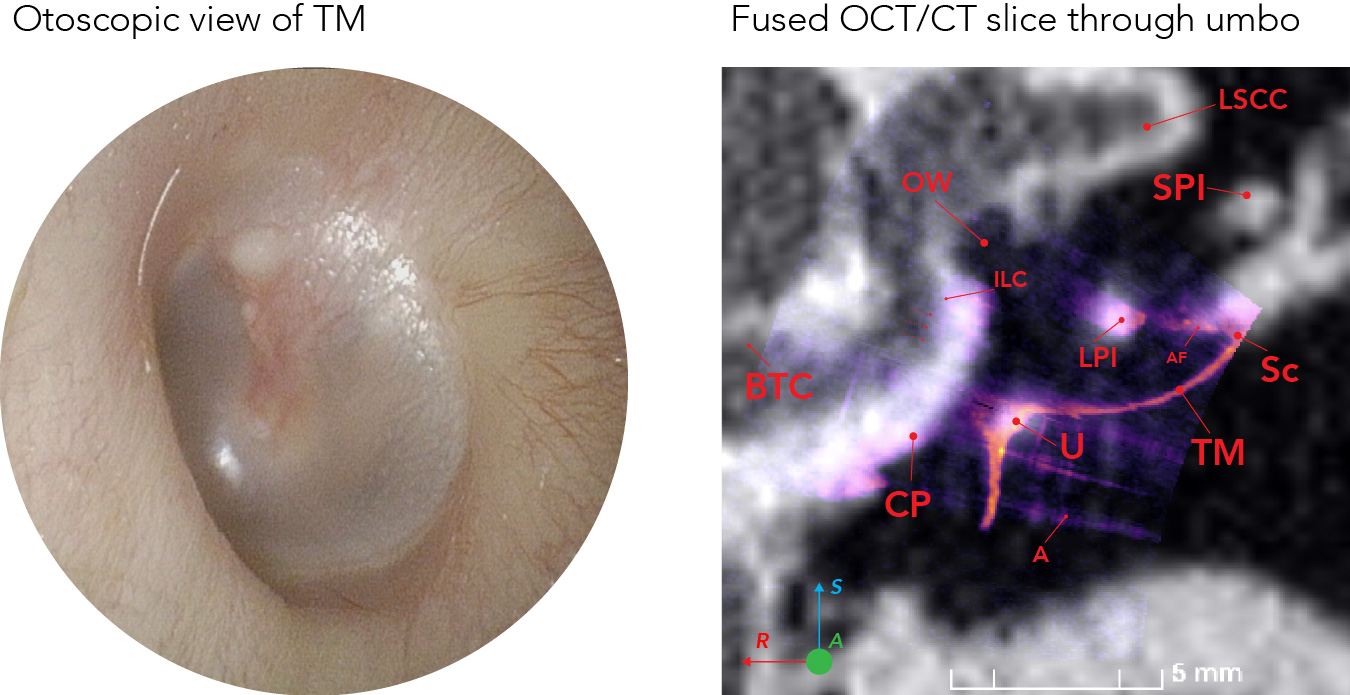
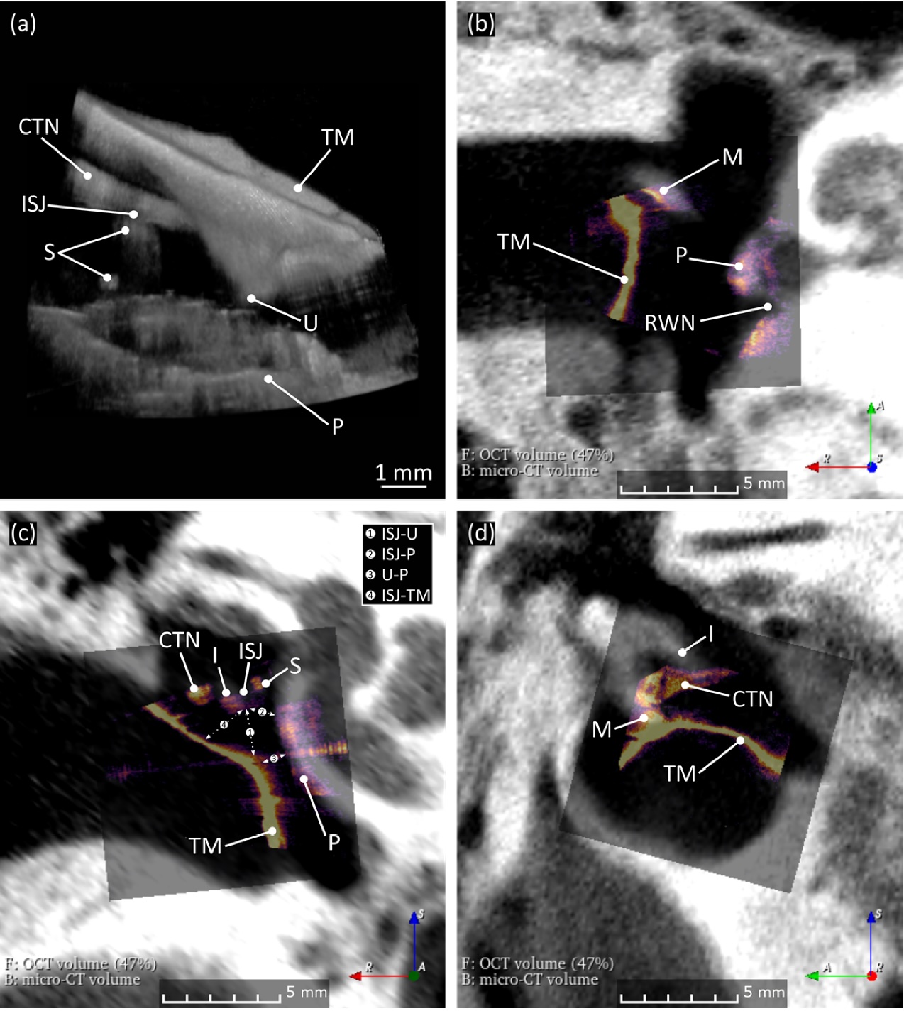
Fusion of Middle Ear Optical Coherence Tomography and Computed Tomography
In this case series including 3 patients, OCT and CT imaging produced complementary diagnostic information, with CT offering unobstructed images of bony anatomy and OCT providing the ability to visualize soft tissue. |
|
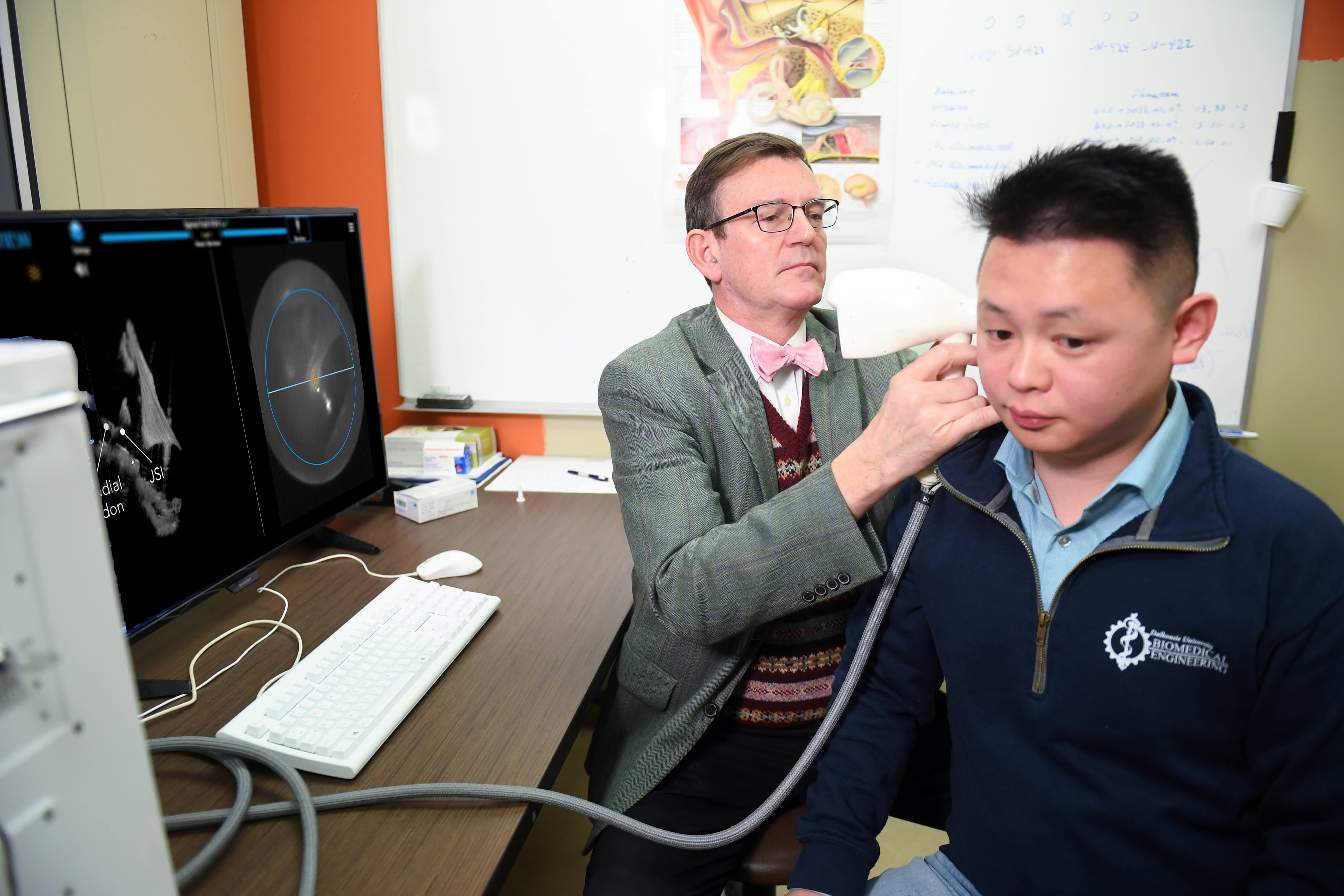
Clinical Applications of Handheld Middle Ear Optical Coherence Tomography (ME-OCT) with Live Volumetric Visualization: Clinical Applications of 4D ME-OCT
A custom-built OCT system enabling continuous 4D imaging of the middle ear for clinical applications such as the Valsalva maneuver, partial ossicular prosthesis (PORP), and cochlear implant (CI) surgeries. |
|
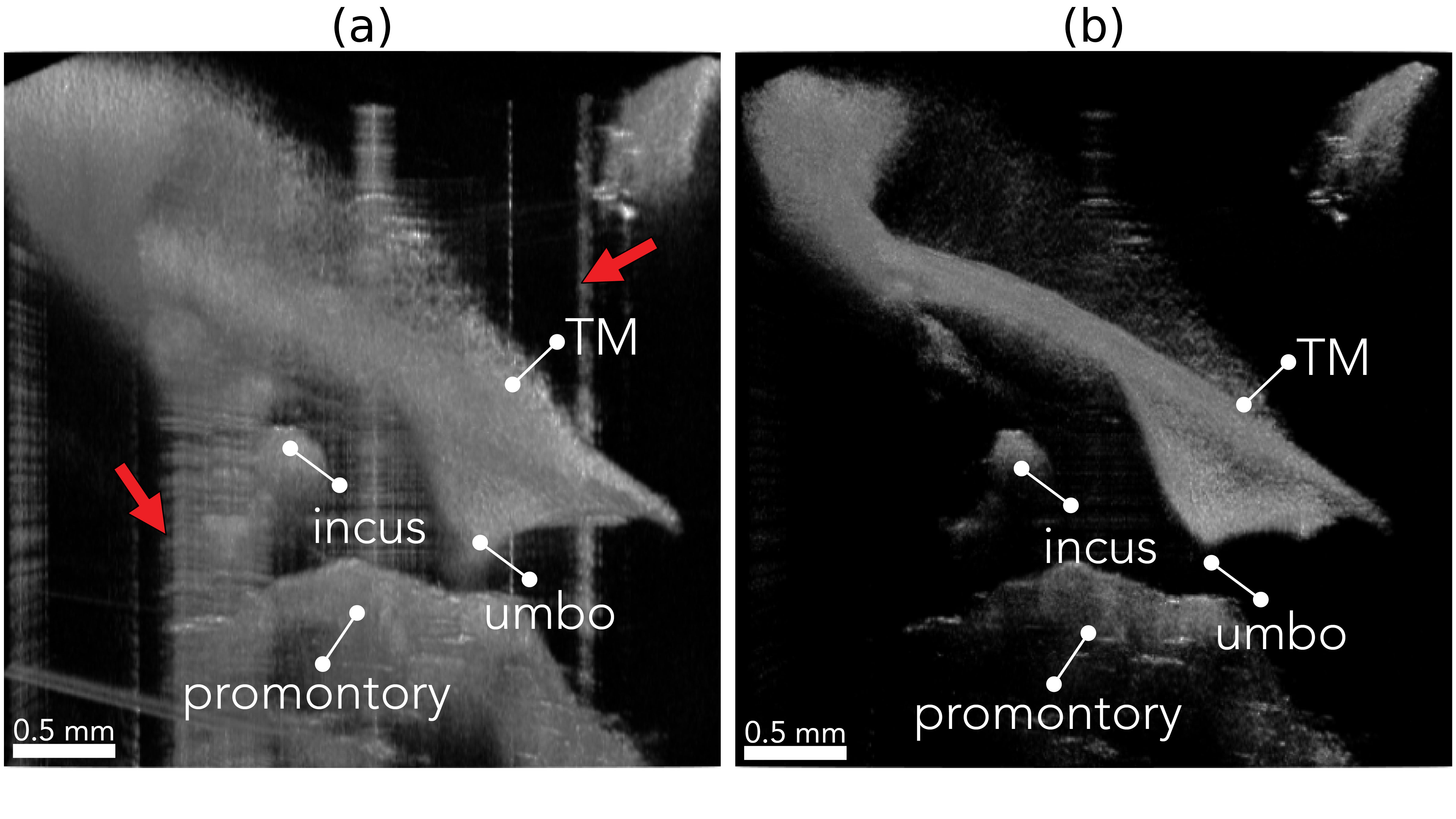
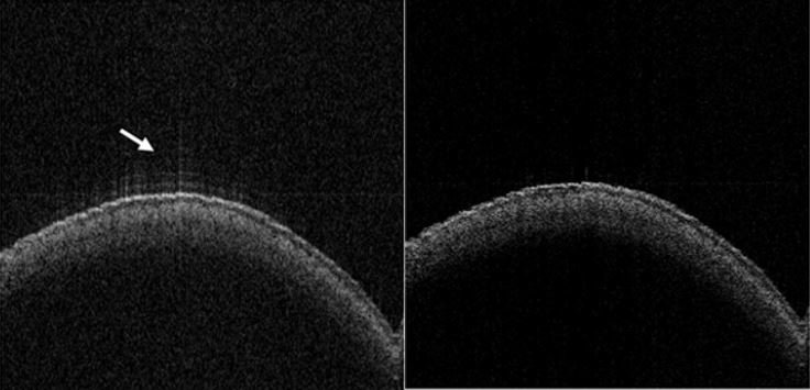
Improved Middle Ear Imaging with Optical Coherence Tomography for Clinical Otology
This thesis introduces two approaches to enhance middle ear OCT visualization: a convolutional basis pursuit framework to reduce sidelobe artifacts and a topical glycerol treatment to improve visualization for postoperative ear. |
|
Geometrically Accurate Real-time Volumetric Visualization of the Middle Ear Using Optical Coherence Tomography
We present a handheld system for live, geometrically accurate volumetric middle ear OCT imaging. Using a discretized spiral scanning (DC-SC) pattern, the system rapidly acquires data and corrects geometric distortions. |

|
Transtympanic Visualization of Cochlear Implant Placement with Optical Coherence Tomography: A Pilot Study
This study evaluates the ability of transtympanic middle ear optical coherence tomography (ME-OCT) to assess placement of cochlear implants (CIs) in situ. |
|
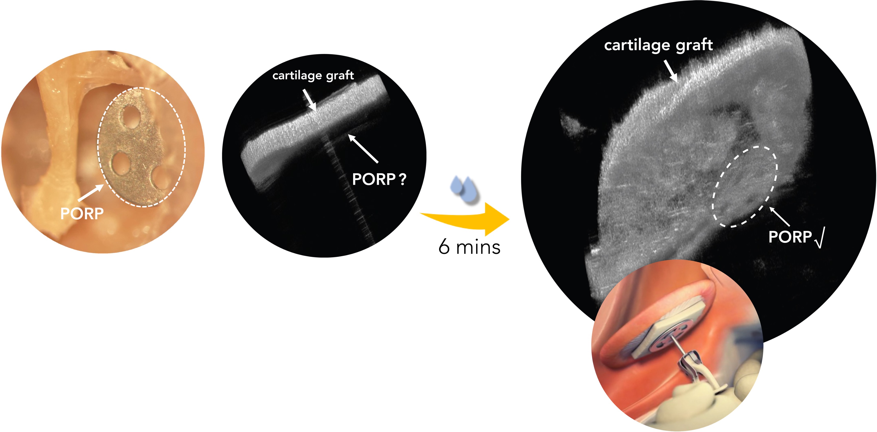
Optical Clearing Agents for Optical Imaging Through Cartilage Tympanoplasties: A Preclinical Feasibility Study
Optical clearing agents (OCAs) can render cartilage tympanoplasty grafts sufficiently transparent to permit visualization of middle ear structures in an operated ear using optical coherence tomography (OCT) imaging. |
|
Convolutional Dictionary Learning for Blind Deconvolution of Optical Coherence Tomography Images
In this study, we demonstrate a sparsity-regularized, complex, blind deconvolution method for removing sidelobe artefacts and stochastic noise from optical coherence tomography (OCT) images. |